

In vitro Zytotoxizitätstest mit direktem Zellkontakt (Mauszelllinie L929)
Kontaktieren Sie unsDer direkte Zellkontakttest ermöglicht eine qualitative Bewertung des zytotoxischen Potenzials eines Prüfgegenstands gemäß ISO 10993-5. Der Test wird durchgeführt, indem der Prüfgegenstand auf eine Monolage von L929-Zellen gelegt wird. Die vom Prüfgegenstand freigesetzten zytotoxischen Substanzen können eine Verformung oder sogar eine Lyse der Zellen unter oder um den Prüfgegenstand herum verursachen, was mikroskopisch beurteilt werden kann.
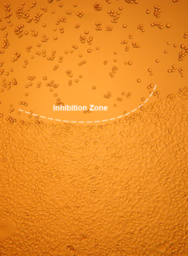
Bewertung der Zytotoxizität durch mikroskopische Auswertung nach direktem Zellkontakt
- Test- und Kontrollproben werden auf die zuvor ausgesäte Zellmonolage aufgebracht, ohne die Integrität der Zellmonolage zu stören, und mindestens 24 Stunden lang bei 37 ± 1°C bebrütet.
- Die Missbildung, Degeneration, Ablösung oder Lyse von Zellen werden als Maß für die Zytotoxizität verwendet. Die biologische Reaktivität wird beschrieben und auf einer Skala von 0 bis 4 bewertet (siehe Tabelle).
- Der Prüfgegenstand erfüllt die Anforderungen des Tests, wenn die Reaktion auf den Prüfgegenstand nicht größer als Grad 2 (leicht reaktiv) ist.


Protokoll |
|
|
Zelllinie |
L929-Zellen (ATCC Nr. CCL1, NCTC-Klon 929 (Bindegewebsmaus), Klon von Stamm L (DSMZ)) |
|
Analyse |
Microscopic evaluation |
|
Inkubationszeit |
24 h bei 37 ± 1°C |
|
Qualitätskontrollen |
Lösungsmittel-Kontrolle: DMEM 10% FBS Negativkontrolle: Polyethylen hoher Dichte (HDPE) Positive Kontrolle: 0,1% Zink-Diethyldithiocarbamat (ZDEC) |
|
Lieferung von Daten |
Zytotoxizität nach Bewertungsskala |
|
Positive Vorhersage |
Der Prüfgegenstand erfüllt die Anforderungen der Prüfung, wenn die Reaktion auf den Prüfgegenstand nicht größer als Grad 2 (leicht reaktiv) ist |
Referenzen
- ISO 10993-5: 2009 “Tests for in vitro cytotoxicity”
- Autian, J. (1977). Toxicological evaluation of biomaterials: Primary acute toxicity screening program. Artif. Organs Vol 1, pp 53 – 60